Differential stains are used to observe bacterial morphology and to divide bacterial cells into distinct groups. Differential staining methods typically require multiple dyes and several staining steps. When used for bacterial identification, differential staining can be combined with other methods.
Gram stain

Figure 1:Gram stain diagram: Gram-positive (left) and Gram-negative (right) bacteria
在微生物学是使用最广泛的污点Gram stain. Based on differences in the structure of the bacterial cell wall, the Gram stain divides bacteria into two major groups: Gram-positive and Gram-negative. Gram-positive cells have a thick layer of peptidoglycan, a polymer made of amino acids and sugars, in the cell wall. Crystal violet binds to peptidoglycan, rendering the cell purple. Gram-negative cells also have peptidoglycan and initially also stain purple. Since the peptidoglycan layer is much thinner, the crystal violet staining is washed out when the cells are exposed to ethanol. They are then stained by the pink counterstain, commonly safranin or fuchsine. Gram-positive cells also take up safranin, but because of the dark purple staining of the cell wall, the lighter pink color can not be observed. The Gram stain is typically the first step in the identification of unknown bacteria. Peptidoglycan is the target of a class of antibiotics called B-lactam antibiotics. While the peptidoglycan layer is thinner in Gram-negative bacteria, these organisms are less susceptible to B-lactam antibiotics as they have an outer membrane, an additional lipid bilayer that surrounds the cell wall. Gram-positive bacteria have a single lipid bilayer inside the cell wall. Like with all staining methods described here, the basic shape and morphology of the bacterium can also be observed in Gram-stained samples.
Acid-fast stain
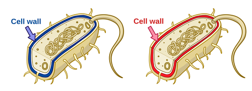
Figure 2:Acid-fast stain diagram: non-acid-fast (left) and acid-fast (right) bacteria
Some bacteria do are not clearly Gram-positive or Gram-negative, includingMycobacteria. These bacteria have mycolic acid in their cell wall. This makes them particularly resistant to decolorization, even with acid alcohols. Inacid-fast stainingprocedures, such as the Ziehl-Neelsen stain, carbol fuchsin stains the cell wall red. Non-acid-fast bacteria lose the red color and are counterstained blue with methylene blue. The acid-fast stain is not as commonly used as the Gram stain. However, since the Mycobacteria include disease-causing organisms, it is an important stain in a clinical setting. The most common example is Mycobacterium tuberculosis, which causes tuberculosis in humans.
Endospore stain
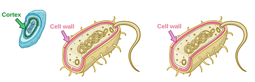
Figure 3:Endospore stain diagram: bacteria with spores (left) and bacteria without spores
Endospores are dormant structures produced by some bacteria when conditions are unfavorable. They can survive without nutrients and are very resistant to extreme temperatures, radiation, and chemical disinfectants. Because of their tough, impermeable exterior, endospores are difficult to stain.Endospore stainingsuch as the Schaeffer-Fulton stain use heat to allow the dye malachite green to penetrate the spores. Similar to the acid-fast stain, the endospores resist decolorization with alcohol and retain the stain. Any bacterial cells (sometimes known as vegetative cells to distinguish them from the spores) are decolorized and counterstained with safranin.